
Check out this Paternoster image:
Image from page 446 of “The animal kingdom : arranged after its organization, forming a natural history of animals, and an introduction to comparative anatomy” (1849)
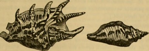
Image by Internet Archive Book Images
Identifier: animalkingdomar00cuvi
Title: The animal kingdom : arranged after its organization, forming a natural history of animals, and an introduction to comparative anatomy
Year: 1849 (1840s)
Authors: Cuvier, Georges, baron, 1769-1832 Metcalf Collection (North Carolina State University) NCRS Blyth, Edward, 1810-1873 Mudie, Robert, 1777-1842 Johnston, George, 1797-1855 Westwood, J. O. (John Obadiah), 1805-1893 Carpenter, William Benjamin, 1813-1885
Subjects: Animals
Publisher: London : Wm. S. Orr and Co., Amen Corner, Paternoster Row
Contributing Library: NCSU Libraries
Digitizing Sponsor: NCSU Libraries
View Book Page: Book Viewer
About This Book: Catalog Entry
View All Images: All Images From Book
Click here to view book online to see this illustration in context in a browseable online version of this book.
Text Appearing Before Image:
ese dismember-ments of the Fusus, Brug., we distinguish the Fasciolariaj Lam., by some oblique and distinct folds on thecolumella, near the origin of the siphon. Turbinella, Lam., are likewise shells with a straight canal, without varices, distinguishable by having [fromthree to five] prominent, compressed, transverse folds, all nearly equal in size, near the centre of the columella,and which approximates them to the conical Volutae: in fact, they only differ by the superior elongation of thesyphonal canal, [and in having an operculum, as well as a thickish epidermis]. The STROMBUSID.E (Sirembus, Linn.)—Comprise the shells with a canal either straight or bent to the right, the external lip of the aperturebecoming, in its maturity, more or less dilated, and always marked with a sinus near the siphonalcanal, whence the head issues when the animal comes out. In the greater number this sinus is at somedistance from the canal. V gcDcra of Montfort.—Ed. GASTEROPODA TUBULIBRANCHIATA. 3G7
Text Appearing After Image:
M. de Lamarck snlidivides Hiosc species into two suoitcnora. His fUromout have tlie outer lip dilated into a wing of more or less expanse, but not dividedinto digitations. The foot is proportion-ably small, and the tentacula support theeyes upon a lateral peduncle larger eventhan the tentaculuin itself. The operculumis horny, lon;^, and narrow, resting upon athin tail. Pteroccraa, Lam., have the mar-pin of the full-grown shell divided into long,slender digitations, varying in number ac-cording to the species. The animal is thesame as in Strombus. Other Strombusidie have the sinus con-tiguous to the siphon. These are the Roi-tellaria, Lam. They have generally a secondcanal mounting up the spire, and formedby the external lip, and by a continuationof the columella. In some of them the lipis digitated. Their animal resembles thatof the MuricidK ; but the operruium is verysmall. Others have merely denticulationson the lip : their canal is long and straight.Others have the margin entire an
Note About Images
Please note that these images are extracted from scanned page images that may have been digitally enhanced for readability – coloration and appearance of these illustrations may not perfectly resemble the original work.
Image automatically curated and published by www.free-mail.co.za – CLICK HERE to learn more about WP Robot for WordPress.
Have you tried: Visiting Port Elizabeth?